3/18/2012

How a Single Gene Mutation Leads to Uncontrolled Obesity


Researchers at Georgetown University Medical Center have revealed how a mutation in a single gene is responsible for the inability of neurons to effectively pass along appetite suppressing signals from the body to the right place in the brain. What results is obesity caused by a voracious appetite. 


Their study, published March 18th on Nature Medicine's website, suggests there might be a way to stimulate expression of that gene to treat obesity caused by uncontrolled eating. 

The research team specifically found that a mutation in the brain-derived neurotrophic factor (Bdnf) gene in mice does not allow brain neurons to effectively pass leptin and insulin chemical signals through the brain. In humans, these hormones, which are released in the body after a person eats, are designed to "tell" the body to stop eating. But if the signals fail to reach correct locations in the hypothalamus, the area in the brain that signals satiety, eating continues. 

"This is the first time protein synthesis in dendrites, tree-like extensions of neurons, has been found to be critical for control of weight," says the study's senior investigator, Baoji Xu, Ph.D., an associate professor of pharmacology and physiology at Georgetown. 

"This discovery may open up novel strategies to help the brain control body weight," he says. 

Xu has long investigated the Bdnf gene. He has found that the gene produces a growth factor that controls communication between neurons. 

For example, he has shown that during development, BDNF is important to the formation and maturation of synapses, the structures that permit neurons to send chemical signals between them. The Bdnf gene generates one short transcript and one long transcript. He discovered that when the long-form Bdnf transcript is absent, the growth factor BDNF is only synthesized in the cell body of a neuron but not in its dendrites. The neuron then produces too many immature synapses, resulting in deficits in learning and memory in mice. 

Xu also found that the mice with the same Bdnf mutation grew to be severely obese. 

Other researchers began to look at the Bdnf gene in humans, and large-scale genome-wide association studies showed Bdnf gene variants are, in fact, linked to obesity. 

But, until this study, no one has been able to describe exactly how BDNF controls body weight. 

Xu's data shows that both leptin and insulin stimulate synthesis of BDNF in neuronal dendrites in order to move their chemical message from one neuron to another through synapses. The intent is to keep the leptin and insulin chemical signals moving along the neuronal highway to the correct brain locations, where the hormones will turn on a program that suppresses appetite. 

"If there is a problem with the Bdnf gene, neurons can't talk to each other, and the leptin and insulin signals are ineffective, and appetite is not modified," Xu says. 

Now that scientists know that BDNF regulates the movement of leptin and insulin signals through brain neurons, the question is whether a faulty transmission line can be repaired. 

One possible strategy would be to produce additional long-form Bdnf transcript using adeno-associated virus-based gene therapy, Xu says. But although this kind of gene therapy has proven to be safe, it is difficult to deliver across the brain blood barrier, he adds. 

"The better approach might be to find a drug that can stimulate Bdnf expression in the hypothalamus," Xu says. "We have opened the door to both new avenues in basic research and clinical therapies, which is very exciting." 

Source: Georgetown University Medical Center [March 18, 2012]

3/15/2012

Believing the Impossible: Research fails to find evidence for the existence of psychic ability


Research failing to find evidence for the existence of psychic ability has been published, following a year of industry debate. The report is a response by a group of independent researchers to the 2011 study from social psychologist Daryl Bem, purporting the existence of precognition -- an ability to perceive future events. 


Professor Chris French (Goldsmiths, University of London), Stuart Ritchie (University of Edinburgh) and Professor Richard Wiseman (University of Hertfordshire) collaborated to accurately replicate Bem's final experiment, and found no evidence for precognition. Their negative results have now been published by open access journal PLoS ONE. 

Their report was rejected by the Journal of Personality and Social Psychology (JPSP), which originally published Bem's findings along with his appeal to independent researchers to attempt replications. 

"Our submission was rejected without being sent for peer review on the basis that the journal has a policy of not publishing replications," said Professor Chris French. "Our paper has opened up the debate on the proper place of replication in the scientific literature." 

In Bem's experiment, after completing a memory test on a list of words, participants were then shown a random selection of half the words from the original list. Results showed that participants were better at remembering the words they were about to be shown, indicating they had reached forward in time to 'practice' those words in the future. 

Within parapsychology, there is a tendency to accept any positive replications but to dismiss failures to replicate if the procedures followed have not been exactly duplicated. 

"We went to great pains to ensure we followed the same procedures as Bem," said Stuart Ritchie. "Using Bem's own computer programme and stats methods, we replicated his experiment three times, at each of our respective campuses, with the same number of participants as the original study." 

"By having our paper published, we hope academic journals and popular media alike will offer the same weight to negative results as given to eye-catching positive results," said Professor Richard Wiseman. 

Source: University of Hertfordshire [March 15, 2012]

3/13/2012

Scientists Produce Eye Structures from Human Blood-Derived Stem Cells


For the first time, scientists at the University of Wisconsin-Madison have made early retina structures containing proliferating neuroretinal progenitor cells using induced pluripotent stem (iPS) cells derived from human blood. 

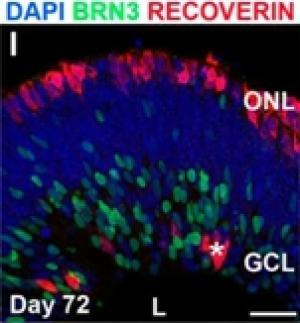
At 72 days, stem cells derived from human blood formed an early retina structure, with specialized cells resembling photoreceptors (red) and ganglion cells (green) located within the outer and inner layers, respectively. Nuclei of cells within the middle layer are shown in blue. These layers are similar to those present during normal human eye development [Credit: University of Wisconsin-Madison]
And in another advance, the retina structures showed the capacity to form layers of cells – as the retina does in normal human development – and these cells possessed the machinery that could allow them to communicate information. (Light-sensitive photoreceptor cells in the retina along the back wall of the eye produce impulses that are ultimately transmitted through the optic nerve and then to the brain, allowing you to see.) Put together, these findings suggest that it is possible to assemble human retinal cells into more complex retinal tissues, all starting from a routine patient blood sample. 

Many applications of laboratory-built human retinal tissues can be envisioned, including using them to test drugs and study degenerative diseases of the retina such as retinitis pigmentosa, a prominent cause of blindness in children and young adults. One day, it may also be possible replace multiple layers of the retina in order to help patients with more widespread retinal damage. 

“We don’t know how far this technology will take us, but the fact that we are able to grow a rudimentary retina structure from a patient’s blood cells is encouraging, not only because it confirms our earlier work using human skin cells, but also because blood as a starting source is convenient to obtain,” says Dr. David Gamm, pediatric ophthalmologist and senior author of the study. “This is a solid step forward.” 

In 2011, the Gamm lab at the UW Waisman Center created structures from the most primitive stage of retinal development using embryonic stem cells and stem cells derived from human skin. While those structures generated the major types of retinal cells, including photoreceptors, they lacked the organization found in more mature retina. 

This time, the team, led by Gamm, Assistant Professor of Ophthalmology and Visual Sciences in the UW School of Medicine and Public Health, and postdoctoral researcher and lead author Dr. Joseph Phillips, used their method to grow retina-like tissue from iPS cells derived from human blood gathered via standard blood draw techniques. 

In their study, about 16 percent of the initial retinal structures developed distinct layers. The outermost layer primarily contained photoreceptors, whereas the middle and inner layers harbored intermediary retinal neurons and ganglion cells, respectively. This particular arrangement of cells is reminiscent of what is found in the back of the eye. Further, work by Dr. Phillips showed that these retinal cells were capable of making synapses, a prerequisite for them to communicate with one another. 

The iPS cells used in the study were generated through collaboration with Cellular Dynamics International (CDI) of Madison, Wis., who pioneered the technique to convert blood cells into iPS cells. CDI scientists extracted a type of blood cell called a T-lymphocyte from the donor sample, and reprogrammed the cells into iPS cells. CDI was founded by UW stem cell pioneer Dr. James Thomson. 

“We were fortunate that CDI shared an interest in our work. Combining our lab’s expertise with that of CDI was critical to the success of this study,” added Dr. Gamm.

Source: University of Wisconsin-Madison [March 13, 2012]

3/09/2012

Healthy aging begins in the womb


Ageing is a complex process involving physical, psychological, and environmental factors. Scientists believe that ageing can be programmed in the womb. One example of stress during pregnancy is the administration of glucocorticoids, i. e. synthetic stress hormones to accelerate fetal lung maturation in premature labor to allow breathing after birth. Could exposure to these stress hormones have an effect on health later in life? 


"We know from previous studies that children whose mothers received glucocorticoids during pregnancy are less stress tolerant and have more concentration problems than their peers," reported Professor Matthias Schwab, the Neurologist heading the project. "Stress signal regulation is permanently disturbed and appears to lead to early ageing, in particular, of the brain", he added. 

The scientists examine several risk groups. One consists of 10-year-olds born at the University Hospital Jena whose mothers had treatment with glucocorticoids during pregnancy. Two other groups are being followed up by psychologists at the University of Leuven in Belgium and include two-year-olds and 25-year-olds whose mothers suffered from stress during pregnancy. 

"A unique group studied at the Academic Medical Centre, University of Amsterdam includes persons born in Holland during the Dutch „Hunger Winter", of 1944/45," reported Prof. Schwab. 

Scientists involved in the project use state-of-the art neuropsychological, neurophysiological, and MRI methods to analyse and compare biological and real age of the brain. The biotechnology companies Life Length in Madrid and Biocrates in Innsbruck measure biological age in chromosomes and screen blood samples to determine metabolic blood markers of age. 

"Environmental factors such as exposure to stress hormones or malnutrition during pregnancy change the readout of genetic information permanently," reported Matthias Platzer, Head of Genome Analysis at the Leibniz Institute for Age Research in Jena. 

"These epigenetic processes significantly change stress sensitivity for the rest of life," explained Prof Schwab. „Increased stress sensitivity makes one vulnerable to age-related diseases such as stroke and depression," he added. 

In experimental studies, researchers aim to determine the period during pregnancy in which the brain is particularly vulnerable to stress. An important aspect of the research also involves studying a group of primates kept in Texas for possible translation of the findings to human subjects. 

Understanding the biology of healthy ageing is desperately needed due to the worldwide ageing population and the economic costs of treating age-related diseases. 

"Environmental influences during life in the womb are early determinants for life-long good health and may be a target for preventive measures against early-ageing and age-related diseases," explained Prof. Schwab.  

Source: Friedrich-Schiller-Universitaet Jena [March 08, 2012]

Twitter Delicious Facebook Digg Stumbleupon Favorites More

 
Design by Free WordPress Themes | Bloggerized by Lasantha - Premium Blogger Themes | Facebook Themes