1/31/2012

Testosterone Makes Us Less Cooperative and More Egocentric


Testosterone makes us overvalue our own opinions at the expense of cooperation, research from the Wellcome Trust Centre for Neuroimaging at UCL (University College London) has found. The findings may have implications for how group decisions are affected by dominant individuals. 


Problem solving in groups can provide benefits over individual decisions as we are able to share our information and expertise. However, there is a tension between cooperation and self-orientated behaviour: although groups might benefit from a collective intelligence, collaborating too closely can lead to an uncritical groupthink, ending in decisions that are bad for all. 

Attempts to understand the biological mechanisms behind group decision making have tended to focus on the factors that promote cooperation, and research has shown that people given a boost of the hormone oxytocin tend to be cooperative. Now, in a study recently published in the journal Proceedings of the Royal Society B, researchers have shown that the hormone testosterone has the opposite effect -- it makes people act less cooperatively and more egocentrically. 

Dr Nick Wright and colleagues at the Wellcome Trust Centre for Neuroimaging at UCL carried out a series of tests using 17 pairs of female volunteers* who had previously never met. The test took place over two days, spaced a week apart. On one of the days, both volunteers in each pair were given a testosterone supplement; on the other day, they were given a placebo. 

During the experiment, both women sat in the same room and viewed their own screen. Both individuals saw exactly the same thing. First, in each trial they were shown two images, one of which contained a high-contrast target -- and their job was to decide individually which image contained the target. 

If their individual choices agreed, they received feedback and moved on to the next trial. However, if they disagreed, they were asked to collaborate and discuss with their partner to reach a joint decision. One of the pair then input this joint decision. 

The researchers found that, as expected, cooperation enabled the group to perform much better than the individuals alone when individuals had received only the placebo. But, when given a testosterone supplement, the benefit of cooperation was markedly reduced. In fact, higher levels of testosterone were associated with individuals behaving egocentrically and deciding in favour of their own selection over their partner's. 

"When we are making decisions in groups, we tread a fine line between cooperation and self-interest: too much cooperation and we may never get our way, but if we are too self-orientated, we are likely to ignore people who have real insight," explains Dr Wright. 

"Our behaviour seems to be moderated by our hormones -- we already know that oxytocin can make us more cooperative, but if this were the only hormone acting on our decision-making in groups, this would make our decisions very skewed. We have shown that, in fact, testosterone also affects our decisions, by making us more egotistical. 

"Most of the time, this allows us to seek the best solution to a problem, but sometimes, too much testosterone can help blind us to other people's views. This can be very significant when we are talking about a dominant individual trying to assert his or her opinion in, say, a jury." 

Testosterone is implicated in a variety of social behaviours. For example, in chimpanzees, levels of testosterone rise ahead of a confrontation or a fight. In female prisoners, studies have found that higher levels of testosterone correlate with increased antisocial behaviour and higher aggression. Researchers believe that such findings reflect a more general role for testosterone in increasing the motivation to dominate others and increase egocentricity. 

Commenting on the findings, Dr John Williams, Head of Neuroscience and Mental Health at the Trust, said: "Cooperating with others has obvious advantages for sharing skills and experience, but we know it doesn't always work, particularly if one alpha male or alpha female dominates the decision making. This result helps us understand at a hormonal level the factors that can disrupt our attempts to work together." 

The Wellcome Trust funded this study. 

*Testosterone is naturally secreted in men and women, and testosterone levels are correlated with important behaviours (e.g. antisocial behaviour) in both men and women. For the size of dose given experimentally, in women this markedly increases their testosterone from its low baseline level. In men, however, the situation is more complicated: men already have high baseline levels of testosterone, so giving such doses will decrease their own production of testosterone, a feedback effect that will act to offset the increase caused by the treatment itself. The researchers therefore used female subjects because giving standard experimental doses causes a straightforward and well-characterised increase in their testosterone levels. 

Source: Wellcome Trust [January 31, 2012]

1/30/2012

Music training has biological impact on aging process


Age-related delays in neural timing are not inevitable and can be avoided or offset with musical training, according to a new study from Northwestern University. The study is the first to provide biological evidence that lifelong musical experience has an impact on the aging process. 


Measuring the automatic brain responses of younger and older musicians and non-musicians to speech sounds, researchers in the Auditory Neuroscience Laboratory discovered that older musicians had a distinct neural timing advantage. 

"The older musicians not only outperformed their older non-musician counterparts, they encoded the sound stimuli as quickly and accurately as the younger non-musicians," said Northwestern neuroscientist Nina Kraus. "This reinforces the idea that how we actively experience sound over the course of our lives has a profound effect on how our nervous system functions." 

Kraus, professor of communication sciences in the School of Communication and professor of neurobiology and physiology in the Weinberg College of Arts and Sciences, is co-author of "Musical experience offsets age-related delays in neural timing" published online in the journal Neurobiology of Aging. 

"These are very interesting and important findings," said Don Caspary, a nationally known researcher on age-related hearing loss at Southern Illinois University School of Medicine. "They support the idea that the brain can be trained to overcome, in part, some age-related hearing loss." 

"The new Northwestern data, with recent animal data from Michael Merzenich and his colleagues at University of California, San Francisco, strongly suggest that intensive training even late in life could improve speech processing in older adults and, as a result, improve their ability to communicate in complex, noisy acoustic environments," Caspary added. 

Previous studies from Kraus' Auditory Neuroscience Laboratory suggest that musical training also offset losses in memory and difficulties hearing speech in noise -- two common complaints of older adults. The lab has been extensively studying the effects of musical experience on brain plasticity across the life span in normal and clinical populations, and in educational settings. 

However, Kraus warns that the current study's findings were not pervasive and do not demonstrate that musician's have a neural timing advantage in every neural response to sound. "Instead, this study showed that musical experience selectively affected the timing of sound elements that are important in distinguishing one consonant from another." 

The automatic neural responses to speech sounds delivered to 87 normal-hearing, native English-speaking adults were measured as they watched a captioned video. "Musician" participants began musical training before age 9 and engaged consistently in musical activities through their lives, while "non-musicians" had three years or less of musical training.  

Source: Northwestern University [January 30, 2012]

1/29/2012

Body clock receptor linked to diabetes in new genetic study


A study published in Nature Genetics today has found new evidence for a link between the body clock hormone melatonin and type 2 diabetes. The study found that people who carry rare genetic mutations in the receptor for melatonin have a much higher risk of type 2 diabetes.

People who carry rare genetic mutations in the receptor for melatonin have a much higher risk of type 2 diabetes, according a study published in Nature Genetics
The findings should help scientists to more accurately assess personal diabetes risk and could lead to the development of personalised treatments.

Previous research has found that people who work night shifts have a higher risk of type 2 diabetes and heart disease. Studies have also found that if volunteers have their sleep disrupted repeatedly for three days, they temporarily develop symptoms of diabetes.

The body's sleep-wake cycle is controlled by the hormone melatonin, which has effects including drowsiness and lowering body temperature. In 2008, a genetic study led by Imperial College London discovered that people with common variations in the gene for MT2, a receptor for melatonin, have a slightly higher risk of type 2 diabetes.

The new study reveals that carrying any of four rare mutations in the MT2 gene increases a person's risk of developing type 2 diabetes six times. The release of insulin, which regulates blood sugar levels, is known to be regulated by melatonin. The researchers suggest that mutations in the MT2 gene may disrupt the link between the body clock and insulin release, leading to abnormal control of blood sugar.

Professor Philippe Froguel, from the School of Public Health at Imperial College London, who led the study, said: "Blood sugar control is one of the many processes regulated by the body's biological clock. This study adds to our understanding of how the gene that carries the blueprint for a key component in the clock can influence people's risk of diabetes.

"We found very rare variants of the MT2 gene that have a much larger effect than more common variants discovered before. Although each mutation is rare, they are common in the sense that everyone has a lot of very rare mutations in their DNA. Cataloguing these mutations will enable us to much more accurately assess a person's risk of disease based on their genetics."

In the study, the Imperial team and their collaborators at several institutions in the UK and France examined the MT2 gene in 7,632 people to look for more unusual variants that have a bigger effect on disease risk. They found 40 variants associated with type 2 diabetes, four of which were very rare and rendered the receptor completely incapable of responding to melatonin. The scientists then confirmed the link with these four variants in an additional sample of 11,854 people.

Professor Froguel and his team analysed each mutation by testing what effect they have on the MT2 receptor in human cells in the lab. The mutations that completely prevented the receptor from working proved to have a very big effect on diabetes risk, suggesting that there is a direct link between MT2 and the disease.

Source: Imperial College London [January 29, 2012]

1/27/2012

Making memories last


Memories in our brains are maintained by connections between neurons called "synapses". But how do these synapses stay strong and keep memories alive for decades? Neuroscientists at the Stowers Institute for Medical Research have discovered a major clue from a study in fruit flies: Hardy, self-copying clusters or oligomers of a synapse protein are an essential ingredient for the formation of long-term memory. 

Drosophila Orb2 plays an important role in the persistence of memory. Upon stimulation, Orb2 (shown in yellow) forms amyloid-like oligomers (shown in red), which are an essential ingredient for the formation of long-term memory [Credit: Illustration: Nicolle Rager Fuller, Sayo-Art]
The finding supports a surprising new theory about memory, and may have a profound impact on explaining other oligomer-linked functions and diseases in the brain, including Alzheimer's disease and prion diseases. 

"Self-sustaining populations of oligomers located at synapses may be the key to the long-term synaptic changes that underlie memory; in fact, our finding hints that oligomers play a wider role in the brain than has been thought," says Kausik Si, Ph.D., an associate investigator at the Stowers Institute, and senior author of the new study, which is published in the January 27, 2012 online issue of the journal Cell. 

Si's investigations in this area began nearly a decade ago during his doctoral research in the Columbia University laboratory of Nobel-winning neuroscientist Eric Kandel. He found that in the sea slug Aplysia californica, which has long been favored by neuroscientists for memory experiments because of its large, easily-studied neurons, a synapse-maintenance protein known as CPEB (Cytoplasmic Polyadenylation Element Binding protein) has an unexpected property. 

A portion of the structure is self-complementary and—much like empty egg cartons—can easily stack up with other copies of itself. CPEB thus exists in neurons partly in the form of oligomers, which increase in number when neuronal synapses strengthen. These oligomers have a hardy resistance to ordinary solvents, and within neurons may be much more stable than single-copy "monomers" of CPEB. They also seem to actively sustain their population by serving as templates for the formation of new oligomers from free monomers in the vicinity. 

CPEB-like proteins exist in all animals, and in brain cells they play a key role in maintaining the production of other synapse-strengthening proteins. Studies by Si and others in the past few years have hinted that CPEB's tendency to oligomerize is not merely incidental, but is indeed essential to its ability to stabilize longer-term memory. "What we've lacked till now are experiments showing this conclusively," Si says. 

In the new study, Si and his colleagues examined a Drosophila fruit fly CPEB protein known as Orb2. Like its counterpart in Aplysia, it forms oligomers within neurons. "We found that these Orb2 oligomers become more numerous in neurons whose synapses are stimulated, and that this increase in oligomers happens near synapses," says lead author Amitabha Majumdar, Ph.D., a postdoctoral researcher in Si's lab. 

The key was to show that the disruption of Orb2 oligomerization on its own impairs Orb2's function in stabilizing memory. Majumdar was able to do this by generating an Orb2 mutant that lacks the normal ability to oligomerize yet maintains a near-normal concentration in neurons. Fruit flies carrying this mutant form of Orb2 lost their ability to form long-term memories. "For the first 24 hours after a memory-forming stimulus, the memory was there, but by 48 hours it was gone, whereas in flies with normal Orb2 the memory persisted," Majumdar says. 

Si and his team are now following up with experiments to determine for how long Orb2 oligomers are needed to keep a memory alive. "We suspect that they need to be continuously present, because they are self-sustaining in a way that Orb2 monomers are not," says Si. 

The team's research also suggests some intriguing possibilities for other areas of neuroscience. This study revealed that Orb2 proteins in the Drosophila nervous system come in a rare, highly oligomerization-prone form (Orb2A) and a much more common, much less oligomerization-prone form (Orb2B). "The rare form seems to be the one that is regulated, and it seems to act like a seed for the initial oligomerization, which pulls in copies of the more abundant form," Si says. "This may turn out to be a basic pattern for functional oligomers." 

The findings may help scientists understand disease-causing oligomers too. Alzheimer's, Parkinson's and Huntington's disease, as well as prion diseases such as Creutzfeldt-Jakob disease, all involve the spread in the brain of apparently toxic oligomers of various proteins. One such protein, strongly implicated in Alzheimer's disease, is amyloid beta; like Orb2 it comes in two forms, the highly oligomerizing amyloid-beta-42 and the relatively inert amyloid-beta-40. Si's work hints at the possibility that oligomer-linked diseases are relatively common in the brain because the brain evolved to be relatively hospitable to CPEB proteins and other functional oligomers, and thus has fewer mechanisms for keeping rogue oligomers under control. 

Source: Stowers Institute for Medical Research [January 27, 2012]

1/24/2012

'Genetic programming': The mathematics of taste


The design of aromas — the flavors of packaged food and drink and the scents of cleaning products, toiletries and other household items — is a multibillion-dollar business. The big flavor companies spend tens of millions of dollars every year on research and development, including a lot of consumer testing. 

[Graphic: Christine Daniloff]
But making sense of taste-test results is difficult. Subjects’ preferences can vary so widely that no clear consensus may emerge. Collecting enough data about each subject would allow flavor companies to filter out some of the inconsistencies, but after about 40 flavor samples, subjects tend to suffer “smell fatigue,” and their discriminations become unreliable. So companies are stuck making decisions on the basis of too little data, much of it contradictory. 

One of the biggest flavor companies in the world has turned to researchers in MIT’s Computer Science and Artificial Intelligence Laboratory (CSAIL) for help. To analyze taste-test results, the CSAIL researchers are using genetic programming, in which mathematical models compete with each other to fit the available data and then cross-pollinate to produce models that are more accurate still. 

The Swiss flavor company Givaudan asked CSAIL principal research scientist Una-May O’Reilly, postdoc Kalyan Veeramachaneni and the University of Antwerp’s Ekaterina Vladislavleva to help interpret the results of tests in which 69 subjects evaluated 36 different combinations of seven basic flavors, assigning each a score according to its olfactory appeal. 

For each subject, O’Reilly and her colleagues randomly generate mathematical functions that predict scores according to the concentrations of different flavors. Each function is assessed according to two criteria: accuracy and simplicity. A function that, for example, predicts a subject’s preferences fairly accurately using a single factor — say, concentration of butter — could prove more useful than one that yields a slightly more accurate prediction but requires a complicated mathematical manipulation of all seven variables. 

After all the functions have been assessed, those that provide poor predictions are winnowed out. Elements of the survivors are randomly recombined to produce a new generation of functions; those are then evaluated for accuracy and simplicity. The whole process is repeated about 30 times, until it converges on a set of functions that accord well with the preferences of a single subject.  

Because O’Reilly and her colleagues’ method produces profiles of individual test subjects’ tastes, it can sort them into distinct groups. It could be, for instance, that test subjects tend to have strong preferences for either cinnamon or nutmeg but not both. By marketing one product to cinnamon lovers and another to nutmeg lovers, a company could do much better than by marketing one product to both. “For every one of these 36 flavors, someone hated it and someone liked it,” O’Reilly says. “If you try to identify a flavor that the whole panel likes, you end up settling for a little bit less.” 

O’Reilly and her colleagues haven’t had an opportunity to empirically determine whether their models correctly predict subjects’ responses to new flavors. So to try to establish their model’s accuracy, they instead built another model. First, they developed a set of mathematical functions that represent subjects’ true taste preferences. Then they showed that, given the limitations of particular test designs, their algorithms could still divine those preferences. Although they developed the model purely to validate their approach, O’Reilly says, flavor researchers were intrigued by the possibility of using it to develop more accurate and efficient test protocols. 

“People have been playing with these [evolutionary] techniques for decades,” says Lee Spector, a professor of computer science at Hampshire College and editor-in-chief of the journal Genetic Programming and Evolvable Machines, where the MIT researchers’ latest paper appears. “One of the reasons that they haven’t made a big splash until recently is that people haven’t really figured out, I think, where they can pay off big.” Taste preference, Spector says, “is a pretty brilliant area in which to apply the evolutionary methods — and it looks as though they’re working, also, so that’s exciting.” 

Author: Larry Hardesty | Source: Massachusetts Institute of Technology [January 24, 2012]

1/23/2012

Lifelong brain-stimulating habits linked to lower Alzheimer’s protein levels


A new study led by researchers at the University of California, Berkeley, provides even more reason for people to read a book or do a puzzle, and to make such activities a lifetime habit. 

PET scans reveal amyloid plaques, which appear as warm colors such as red and orange. The middle scan is from a person with no symptoms of cognitive problems, but with evident levels of amyloid plaque in the brain [Credit: Susan Landau and William Jagust]
Brain scans revealed that people with no symptoms of Alzheimer’s who engaged in cognitively stimulating activities throughout their lives had fewer deposits of beta-amyloid, a destructive protein that is the hallmark of the disease. 

While previous research has suggested that engaging in mentally stimulating activities – such as reading, writing and playing games – may help stave off Alzheimer’s later in life, this new study identifies the biological target at play. This discovery could  guide future research into effective prevention strategies. 

“These findings point to a new way of thinking about how cognitive engagement throughout life affects the brain,” said study principal investigator Dr. William Jagust,  a professor with joint appointments at UC Berkeley’s Helen Wills Neuroscience Institute, the School of Public Health and Lawrence Berkeley National Laboratory. “Rather than simply providing resistance to Alzheimer’s, brain-stimulating activities may affect a primary pathological process in the disease. This suggests that cognitive therapies could have significant disease-modifying treatment benefits if applied early enough, before symptoms appear.” 

An estimated 5.4 million Americans live with Alzheimer’s disease, but the numbers are growing as baby boomers age. Between 2000 and 2008, deaths from Alzheimer’s increased 66 percent, making it the sixth-leading killer in the country. There is currently no cure, but a draft of the first-ever National Alzheimer’s Plan, released this week, revealed that the U.S. government is aiming for effective Alzheimer’s treatments by 2025. 

The new study, to be published Monday, Jan. 23, in the Archives of Neurology, puts the spotlight on amyloid – protein fibers folded into tangled plaques that accumulate in the brain. Beta-amyloid is considered the top suspect in the pathology of Alzheimer’s disease, so finding a way to reduce its development has become a major new direction of research. 

A molecular model of amyloid protein fibrils. Formed when mis-folded proteins self-assemble into fibrous sheet structures, they are found in the brains of sufferers of Alzheimer's disease [Credit: Susan Landau and William Jagust]
The researchers note that the buildup of amyloid can also be influenced by genes and aging – one-third of people age 60 and over have some amyloid deposits in their brain – but how much reading and writing one does is under each individual’s control. 

“This is the first time cognitive activity level has been related to amyloid buildup in the brain,” said study lead author Susan Landau, research scientist at the Helen Wills Neuroscience Institute and the Berkeley Lab. “Amyloid probably starts accumulating many years before symptoms appear. So it’s possible that by the time you have symptoms of Alzheimer’s, like memory problems, there is little that can be done to stop disease progression. The time for intervention may be much sooner, which is why we’re trying to identify whether lifestyle factors might be related to the earliest possible changes.” 

The researchers asked 65 healthy, cognitively normal adults aged 60 and over (average age was 76) to rate how frequently they participated in such mentally engaging activities as going to the library, reading books or newspapers, and writing letters or email. The questions focused on various points in life from age 6 to the present. 

The participants took part in extensive neuropsychological testing to assess memory and other cognitive functions, and received positron emission tomography (PET) scans at the Berkeley Lab using a new tracer called Pittsburgh Compound B that was developed to visualize amyloid. The results of the brain scans of healthy older individuals with various levels of lifetime cognitive activity were compared with those of 10 patients diagnosed with Alzheimer’s disease and 11 healthy people in their 20s. 

The researchers found a significant association between higher levels of cognitive activity over a lifetime and lower levels of beta-amyloid in the PET scans. They analyzed the impact of other factors such as memory function, physical activity, self-rated memory ability, level of education and gender, and found that lifelong cognitive engagement was independently linked to amyloid deposition. 

Notably, the researchers did not find a strong connection between amyloid deposition and levels of current cognitive activity alone. 

“What our data suggests is that a whole lifetime of engaging in these activities has a bigger effect than being cognitively active just in older age,” said Landau. 

The researchers are careful to point out that the study does not negate the benefits of kicking up brain activity in later years. 

“There is no downside to cognitive activity. It can only be beneficial, even if for reasons other than reducing amyloid in the brain, including social stimulation and empowerment,” said Jagust. “And actually, cognitive activity late in life may well turn out to be beneficial for reducing amyloid. We just haven’t found that connection yet.” 

Other study authors include researchers from UC San Francisco’s Memory and Aging Center and Department of Neurology, and Rush University Medical Center’s Alzheimer’s Disease Center in Chicago. 

The National Institutes of Health and the Alzheimer’s Association helped support this research. 

Author: Sarah Yang | Source: UC Berkeley [January 23, 2012]

1/22/2012

The price of your soul: How the brain decides whether to 'sell out'


An Emory University neuro-imaging study shows that personal values that people refuse to disavow, even when offered cash to do so, are processed differently in the brain than those values that are willingly sold. 


"Our experiment found that the realm of the sacred – whether it's a strong religious belief, a national identity or a code of ethics – is a distinct cognitive process," says Gregory Berns, director of the Center for Neuropolicy at Emory University and lead author of the study. The results were published in Philosophical Transactions of the Royal Society. 

Sacred values prompt greater activation of an area of the brain associated with rules-based, right-or-wrong thought processes, the study showed, as opposed to the regions linked to processing of costs-versus-benefits. 

Berns headed a team that included economists and information scientists from Emory University, a psychologist from the New School for Social Research and anthropologists from the Institute Jean Nicod in Paris, France. The research was funded by the U.S. Office of Naval Research, the Air Force Office of Scientific Research and the National Science Foundation. 

"We've come up with a method to start answering scientific questions about how people make decisions involving sacred values, and that has major implications if you want to better understand what influences human behavior across countries and cultures," Berns says. "We are seeing how fundamental cultural values are represented in the brain." 

The researchers used functional magnetic resonance imaging (fMRI) to record the brain responses of 32 U.S. adults during key phases of an experiment. In the first phase, participants were shown statements ranging from the mundane, such as "You are a tea drinker," to hot-button issues such "You support gay marriage" and "You are Pro-Life." Each of the 62 statements had a contradictory pair, such as "You are Pro-Choice," and the participants had to choose one of each pair. 

At the end of the experiment, participants were given the option of auctioning their personal statements: Disavowing their previous choices for actual money. The participants could earn as much as $100 per statement by simply agreeing to sign a document stating the opposite of what they believed. They could choose to opt out of the auction for statements they valued highly.  

"We used the auction as a measure of integrity for specific statements," Berns explains. "If a person refused to take money to change a statement, then we considered that value to be personally sacred to them. But if they took money, then we considered that they had low integrity for that statement and that it wasn't sacred." 

The brain imaging data showed a strong correlation between sacred values and activation of the neural systems associated with evaluating rights and wrongs (the left temporoparietal junction) and semantic rule retrieval (the left ventrolateral prefrontal cortex), but not with systems associated with reward. 

"Most public policy is based on offering people incentives and disincentives," Berns says. "Our findings indicate that it's unreasonable to think that a policy based on costs-and-benefits analysis will influence people's behavior when it comes to their sacred personal values, because they are processed in an entirely different brain system than incentives." 

Research participants who reported more active affiliations with organizations, such as churches, sports teams, musical groups and environmental clubs, had stronger brain activity in the same brain regions that correlated to sacred values. "Organized groups may instill values more strongly through the use of rules and social norms," Berns says. 

The experiment also found activation in the amygdala region, a brain region associated with emotional reactions, but only in cases where participants refused to take cash to state the opposite of what they believe. "Those statements represent the most repugnant items to the individual," Berns says, "and would be expected to provoke the most arousal, which is consistent with the idea that when sacred values are violated, that induces moral outrage." 

The study is part of a special issue of the Philosophical Transactions of the Royal Society, titled "The Biology of Cultural Conflict." Berns edited the special issue, which brings together a dozen articles on the culture of neuroscience, including differences in the neural processing of people on the opposing sides of conflict, from U.S. Democrats and Republicans to Arabs and Israelis. 

"As culture changes, it affects our brains, and as our brains change, that affects our culture. You can't separate the two," Berns says. "We now have the means to start understanding this relationship, and that's putting the relatively new field of cultural neuroscience onto the global stage." 

Future conflicts over politics and religion will likely play out biologically, Berns says. Some cultures will choose to change their biology, and in the process, change their culture, he notes. He cites the battles over women's reproductive rights and gay marriage as ongoing examples. 

Source: Emory University [January 22, 2012]

1/19/2012

Genetic study offers clues to how intelligence changes through life


Scientists have estimated for the first time the extent to which genes determine changes in intelligence across the human life course. The study found that genetic factors may account for about 24 per cent of changes in intelligence between childhood and old age. The findings also suggest that many of the genes that affect intelligence in childhood also influence intelligence in old age, according to the study published in Nature. The study, by researchers at the Universities of Edinburgh and Queensland and Aberdeen suggests that the largest influence on changes in intelligence is probably environmental. 

DNA from cool angle [Credit: © Dmitry Sunagatov/Fotolia.com]
Identifying genetic influences on intelligence could help us to understand the relationship between knowledge and problem solving and an individual’s outcomes in life, and especially to understand why some people age better than others in terms of intelligence. The researchers combined DNA analysis with data from people who took intelligence tests aged 11 and again aged 65 to 79. The scientists examined more than half a million genetic markers in about 2,000 people to work out how genetically similar they were, even though they were not related. 

The new findings were made possible because Scotland has a rich source of cognitive test data. In June 1932 and June 1947. Intelligence tests were carried out on almost all children born in Scotland in 1921 and 1936, respectively. For the present study, about 2000 of these people were traced and re-tested in old age. 

Professor Ian Deary of the University of Edinburgh’s Centre for Cognitive Ageing and Cognitive Epidemiology, said: “Until now, we have not had an estimate of how much genetic differences affect how intelligence changes across a lifetime. These new findings were possible because our research teams were able to combine a range of valuable resources.    The results partly explain why some people’s brains age better than others. We are careful to suggest that our estimates do not have conventional statistical significance, but they are nevertheless useful because such estimates have been unavailable to date.”  

Professor Peter Visscher of the University of Queensland, said: “Unique data and new genome technologies combined with novel analysis methods allowed us to tackle questions that were not answerable before. The results also strongly suggest how important the environment is helping us to stay sharp as we age. Neither the specific genetic nor environmental factors were identified in this research. Our results provide the warrant for others and ourselves to search for those.” 

Professor Paul Haggarty from the University of Aberdeen Rowett Institute of Nutrition and Health said: "The extensive historical information collected in the Aberdeen and Lothian cohorts included in this study will also make them particularly useful when trying to track down the precise way in which genes and the environment interact to influence cognition throughout life." 

Professor Emeritus of Mental Health at the University of Aberdeen, Lawrence Whalley said: "The Nature research report is a milestone in a long-term research programme that began in Aberdeen in 1997 and was expanded to Edinburgh in 2000. This research collaboration between the universities of Aberdeen and Edinburgh has yielded over 100 joint scientific papers that have added considerably to our understanding of mental decline in old age, and the beginnings of dementia. 

“In this paper, our already substantial collaboration was strengthened by novel methods of data analysis developed by a team in Australia. Using unique Aberdeen and Edinburgh data, with our Australian colleagues we were able to tease apart the overall effects of genes and the environment on causes and courses of mental decline. These findings will encourage others working with complex diseases of old age to apply the same methods to their findings from long-term ageing research." 

Source: University of Aberdeen [January 19, 2012]

1/18/2012

Biologists a Step Nearer to Solving the Parkinson's Conundrum


Scientists at the University of York have made a significant step forward in isolating the cause of Parkinson's disease in younger adults. 

Parkinson's disease occurs when some nerve cells in a part of the brain called the substantia nigra die or become impaired. Normally, these cells produce dopamine - a vital chemical that allows smooth, coordinated function of the body's muscles and movements [Credit: iStockphoto/Martí Sáiz]
Research by a team in the University's Department of Biology found evidence that movement disorders, including tremor and slowness of movement (bradykinesia), associated with Parkinson's disease (PD) may be due to a defect in energy production in the nervous system. The advance may help to identify young adults who may be susceptible to the disease. 

Parkinson's, the second most common form of neurodegenerative disease, principally affects people aged over 60, but some forms -- known as juvenile PD -- usually start in the 30-40 age group. One in 20 people diagnosed with Parkinson's are under 40 and such early onset PD is often inherited. Previous research has identified the genes which cause the disease and found them to be linked in a common pathway to failure of the mitochondria -- the power source within each cell. 

In the latest research, part-funded by leading research charity Parkinson's UK and published in Human Molecular Genetics, scientists at York studied the effect that parkin, one of the genes which cause juvenile PD, has on the larva of the fruit fly, Drosophila. 

They discovered that parkin faithfully models the locomotory defects of PD with a marked reduction in speed, and slower muscle contractions, reminiscent of bradykinesia. 

Dr Chris Elliott, who led the study, said: "Our experimental evidence confirmed that this was due to a defect in the nervous system. This was important because previous work had suggested a big impact on the muscles, but PD is associated with neuronal failure." 

The research team, which included undergraduate and postgraduate students, found that oxygen consumption and the production of ATP (the chief supply of energy inside cells) were drastically reduced. In response, lactate was increased. 

The researchers also discovered that parkin larvae showed oxidative stress due to high levels of reactive oxygen species (ROS; also known as free radicals, such as hydrogen peroxide) which have been suggested as a key component of PD. The study suggested that relieving the ROS had only a marginal effect on mitigating slowed locomotion. 

Dr Elliott added: "These findings show drastic failure in energy production by parkin larvae, and suggest that biochemicals related to lactate may be worth investigating as biomarkers for the progress of PD. 

"We believe that the larval bradykinesia is a consequence of neuronal energy deficit, which leads to failure in neural communication. Oxidative stress is a consequence, rather than cause, of PD." 

Dr Kieran Breen, Director of Research and Innovation at Parkinson's UK, comments: "This study shows just how vital models like the fruit fly are in helping us understand what happens to the nerve cells that are affected in Parkinson's. 

"We already knew that mitochondria were important in Parkinson's but this research suggests that mitochondrial problems may be the root cause of the problems that lead to nerve cell death. 

"So finding ways to protect and enhance the mitochondria may be the key to treatments that can slow or even stop Parkinson's in its tracks." 

Source: University of York [January 16, 2012]

Scientists identify gene crucial to normal development of lungs and brain


Scientists at the Salk Institute for Biological Studies have identified a gene that tells cells to develop multiple cilia, tiny hair-like structures that move fluids through the lungs and brain. The finding may help scientists generate new therapies that use stem cells to replace damaged tissues in the lung and other organs. 

The Salk researchers discovered the master gene that tells cells to develop multiple hair-like cilia, such as those seen in pink in this false-colored electron microscope image of the surface of a frog embryo [Credit: Courtesy of the Salk Institute for Biological Studies; Chris Kintner, Molecular Neurobiology Laboratory, and Matthew Joens and James Fitzpatrick, Waitt Advanced Biophotonics Center]
"Cells with multiple cilia play a number of important roles, including moving fluids through the respiratory tract, brain and spinal cord," says Christopher R. Kintner, a professor in Salk's Molecular Neurobiology Laboratory, who led the research. "Knowing the gene that instructs cells to develop multiple cilia helps us understand how we might coax stem cells into developing into this type of cell, which we could then use to repair damaged tissue." 

The findings of the research, which was supported by the National Institutes of Health and Salk's Innovation Grants Program, were published in the January 8 online issue of Nature Cell Biology. 

Kintner and his collaborator, Jennifer Stubbs, a scientist now at Pathway Genomics, a San Diego biotech company, along with Eszter Vladar, a postdoctoral fellow in the laboratory of Jeffery Axelrod, at the Stanford University School of Medicine, made their discovery by initially studying the embryos of African clawed frogs (Xenopus laevis). 

Multiciliate cells form on the outside of the embryos, making them easy to study, and the genetic mechanisms that direct the frog cells to develop multiple cilia are likely similar to those of humans. 

Humans and other organisms inherited cilia from our single-celled primordial ancestors that used these beating structures as a form of propulsion. Most cells in our body project a single, non-moving cilium used as a tiny antenna for detecting chemical and physical stimuli. But certain specialized tissues require cells with 100 to 200 moving cilia that beat in concert to move fluids through the body. 

These cells aid in pushing cerebrospinal fluid through the brain and spinal cord, helping to circulate and replenish this fluid. In the respiratory system, the cilia push mucus that traps dust, pathogens and other foreign matter from the lung up into the trachea, helping prevent infections. 

In a previous study, published in Nature Genetics, Kintner and Stubbs identified a protein, FoxJ1, that promoted the formation of a single moving cilium. What remained unclear is how certain cells activate FoxJ1 in a way that leads to the formation of hundreds of motile cilia per cell. 

In their new study, Kintner and his collaborators identified a gene that produces a second protein, which they dubbed "multicilin," that tells cells to develop multiple cilia. When cells are exposed to multicilin, their genetic mechanisms for developing multiple cilia are activated. In a developing embryo, the protein instructs certain stem cells that will line the lungs, kidney and skin to develop into multiciliate cells. 

When the researchers inhibited multicilin's action, the frogs' skin and kidney failed to form multiciliate cells. The scientists also found that multicilin is both necessary and sufficient to instruct the development of multiple cilia in cells that line the airways of mice. 

"This means that multicilin directs the development of these cells in a number of different organs," Kintner says. "How multiciliate cells develop had been a mystery, but this fills in a big piece of the puzzle." 

Kintner notes that patients with respiratory diseases such as chronic asthma, emphysema and cystic fibrosis often suffer from lung infections, which may result from damage to the ciliated cells that move protective mucus out of the airways. In the future, stem cell therapies might replace those damage cells with new ciliated cells, but first scientists need to know how to guide stem cells along a pathway into multiciliate cells. 

"Our findings suggest that multicilin could be central to differentiating stem cells into replacement cells," Kintner says. "It's a necessary step in developing such therapies." 

Source: Salk Institute [January 11, 2012]

Moderate marijuana use doesn’t damage lung function


Occasional and low cumulative marijuana use is not associated with adverse effects on pulmonary function, a new study has found. 


Exposure to tobacco smoke causes lung damage with clinical consequences that include respiratory symptoms, chronic obstructive pulmonary disease, and lung cancer. 

Mark J. Pletcher and his colleagues from the University of California examined associations between marijuana, both current and lifetime exposure, and pulmonary function. 

The Coronary Artery Risk Development in Young Adults (CARDIA) study is a longitudinal study collecting repeated measurements of pulmonary function and smoking over 20 years (March 1985-August 2006) in a group of 5,115 men and women in 4 U. S. cities. 

Lifetime exposure to marijuana joints was expressed in joint-years, with 1 joint-year of exposure equivalent to smoking 365 joints or filled pipe bowls. Lung function was assessed by the measurements of forced expiratory volume in the first second of expiration (FEV1) and forced vital capacity (FVC), with lower measures corresponding to poorer lung function. 

More than half of participants reported current marijuana smoking, tobacco smoking, or both at 1 or more examinations. The median intensity of tobacco use in tobacco smokers was substantially higher - 8-9 cigarettes per day, than the median intensity of marijuana use in marijuana smokers -2-3 episodes in the last 30 days. 

In fully adjusted models that considered 4-level categorizations of current and lifetime exposure to tobacco and marijuana, tobacco smoking was associated with a lower FEV1 and current smoking with a lower FVC. 

In contrast, exposure to marijuana was associated with higher FVC and lifetime exposure with higher FEV1. At low lifetime exposure levels, increasing marijuana use was associated with an increase in both FEV1 and FVC. 

"With up to 7 joint-years of life-time exposure e. g., 1 joint a day for 7 years or 1 joint/week for 49 years, we found no evidence that increasing exposure to marijuana adversely affects pulmonary function," the researchers said. 

At more than 10 joint-years of lifetime exposure, there was a nonsignificant decline in FEV1, but there was a significant decline in FEV1 at more than 20 episodes of marijuana use per month. 

"Marijuana may have beneficial effects on pain control, appetite, mood, and management of other chronic symptoms. Our findings suggest that occasional use of marijuana for these or other purposes may not be associated with adverse consequences on pulmonary function," they said. 

"It is more difficult to estimate the potential effects of regular heavy use, because this pattern of use is relatively rare in our study sample; however, our findings do suggest an accelerated decline in pulmonary function with heavy use and a resulting need for caution and moderation when marijuana use is considered," the researchers added. 

The study has been recently published in JAMA. 

Source: Top News [January 11, 2012]

1/17/2012

Permafrost bacteria may slow down aging


A hardy type of bacteria recently discovered in the permafrost of Siberia could help slow down the ageing process, Russian scientists claimed on Tuesday. 


The species of bacteria -- given the name Bacillius F -- was found in laboratory tests to have shown signs of slowing down the process of ageing on mice, the Russian Academy of Sciences (RAN) said. 

The Siberian branch of the RAN said Bacillius F lags 3 million years behind similar bacteria in evolutionary terms, according to the characteristics of proteins and some other factors. 

"Taking into consideration the unusual living environment, one can only marvel at the resilience of these bacteria," it said. 

It added that the organisms found in Russia's northern region of Yakutia -- home to the coldest inhabited area on the planet -- reproduce at just 5 degrees Celsius. 

"We just thought: since the bacteria were found in the permafrost where they were successfully preserved they will possibly have mechanisms of retaining viability," added Nadezhda Mironova, senior research scientist at the Institute of Chemical Biology and Fundamental Medicine of the Russian Academy of Sciences. 

"This is what happened," she was quoted as saying. 

Injections of the bacteria into mice have helped boost the natural defences of the animals as they grew older. 

"Bacillius F injections have favourably affected the quality of being of the aging animals," the Russian scientists said. 

"First and foremost, this concerns immunity and the speed of its activation." 

Experiments have shown that metabolism in the tested mice have increased by 20 to 30 percent, the scientists said, adding that the bacterium may also reduce instances of senile blindness but not the emergence of tumours. 

The Russian Academy of Sciences did not say how many mice were tested, adding more animals were needed for the experiments to be more reliable. The mice from a test group lived longer than those in a control group however, it said, calling the results "impressive." 

Source: AFP [January 17, 2012]

1/12/2012

Opioids Erase Memory Traces of Pain


A team of researchers at the MedUni Vienna's Department of Neurophysiology (Centre for Brain Research) has discovered a previously unknown effect of opioids: the study, which has now been published in the journal Science and was led by Ruth Drdla-Schutting and Jurgen Sandkuhler, shows that opioids not only temporarily relieve pain, but at the right dose can also erase memory traces of pain in the spinal cord and therefore eliminate a key cause of chronic pain. 


The scientists recreated a surgical procedure in vivo in which pain fibres were stimulated under controlled conditions. 

Says Sandkuhler: "Although deep anaesthesia prevents any sensations of pain, we were able to reserve long-term synaptic potentiation in the spinal cord. Despite anaesthesia, there appears to be a memory trace for pain and a pain amplifier has engaged." High doses of intravenous opioids over the course of an hour -- normally opioids are delivered at moderate doses over a longer period -- were able to completely resolve the potentiation. Says Sandkuhler: "The memory trace for pain was therefore deleted again and the pain amplifier switched off." 

The memory trace, as it is termed, is triggered by a variety of mechanisms, including the potentiation of signal transmission at the contact points (synapses) between the nerve cells. This is known as long-term synaptic potentiation. This pain memory can result in the sensation of amplified pain lasting much longer than the actual cause of the pain, even leading to a condition known as chronic pain syndrome. 

A paradigm shift in pain therapy? 

The project, which is sponsored by the Vienna Fund for Science, Research and Technology (WWTF), is currently investigating how this discovery can be put to use in clinical settings. To this end, test subjects or patients with pain syndrome are being given a high dose of an opioid over a period of 60 minutes. 

"If our approach turns out to be effective under clinical conditions, this would herald a paradigm shift in pain therapy. It would mean moving away from the temporary, purely symptom-based pain therapy to a long-term removal of the cause of pain based on pain mechanisms using opioids." 

The effect of opioids (morphine or morphine-like substances) is based on their ability to bind to specific binding sites, known as µ-opiate receptors (MOR) which are found on nerve cells and which process pain-related information. Until now, it has been assumed that opioids are only able to alleviate pain while they are bound to the MOR and therefore suppress stimulation in the pain-processing system. Says Drdla-Schutting: "As soon as the medication is stopped, the pain-relieving effect disappears too." In clinical practice, opioids are therefore given continuously in moderate doses in order to achieve permanent binding to the MOR. This may relieve pain very effectively, but its cause cannot be eliminated. The new, high-dose, short-term therapy with opioids, on the other hand, causes a reversal of cellular changes that play an important role in pain memories, therefore possibly eliminating one of the causes of chronic pain. 

Source: Medical University of Vienna [January 12, 2012]

1/10/2012

Powerful people overestimate their height


The psychological experience of power makes people feel taller than they are, according to research by ILR School associate professor of organizational behavior Jack Goncalo and a Washington University colleague. 


"Although a great deal of research has shown that physically imposing individuals are more likely to acquire power, this work is the first to show that the powerful may actually feel taller than they are," Goncalo and Michelle Duguid write in an upcoming issue of Psychological Science. 

So a 5-foot-4-inch woman might actually sprout an inch or two in her own mind when she's having an empowered moment. In other words, there is actually a physical experience that goes along with feeling powerful. 

Three experiments with 266 American men and women confirmed for Goncalo and Duguid that there is a relationship between feelings of power and one's self-perception of height. 

"Using different manipulations of power and measures of perceived height, we found that people literally perceived themselves as taller when they occupied a more powerful position," they write. 

Is that perhaps why, Goncalo and Duguid wonder, BP Chairman Carl-Henric Svanberg referred to Gulf of Mexico oil spill victims as "small people?" 

Research -- included assigning one's height to a video game avatar --established a starting point for exploring reciprocity between the psychological and physical experiences of power, Goncalo said. 

He added that the research begs a number of questions: Do short people attempt to capture power by physically elevating themselves above others? Would it be possible to psychologically empower people by giving them an office on the top floor? Can feeling powerful make leaders less able to feel empathy and relate to the "little people" because they literally feel bigger? Is it possible that BP's CEO gave us an insider's view on the experience of power? 

Maybe the powerful really do feel bigger than the rest of us. 

Author: Mary Catt | Source: Cornell University [January 09, 2012]

1/09/2012

Getting Cancer Cells to Swallow Poison


Honing chemotherapy delivery to cancer cells is a challenge for many researchers. Getting the cancer cells to take the chemotherapy "bait" is a greater challenge. But perhaps such a challenge has not been met with greater success than by the nanotechnology research team of Omid Farokhzad, MD, Brigham and Women's Hospital (BWH) Department of Anesthesiology Perioperative and Pain Medicine and Research. 

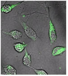
Ligand-nanoparticle components (in green) targeting and binding to cells [Credit: Image courtesy of Brigham and Women's Hospital]
In their latest study with researchers from Massachusetts Institute of Technology (MIT) and Massachusetts General Hospital, the BWH team created a drug delivery system that is able to effectively deliver a tremendous amount of chemotherapeutic drugs to prostate cancer cells. 

The study is electronically published in the January 3, 2012 issue of ACS Nano. 

The process involved is akin to building and equipping a car with the finest features, adding a passenger (in this case the cancer drug), and sending it off to its destination (in this case the cancer cell). 

To design the "vehicle," researchers used a selection strategy developed by Farokhzad's team that allowed them to essentially select for ligands (molecules that bind to the cell surface) that could specifically target prostate cancer cells. The researchers then attached nanoparticles containing chemotherapy, in this case docetaxel, to these hand-picked ligands. 

To understand Farokhzad's selection strategy, one must understand ligand behavior. While most ligands mainly have the ability to bind to cells, the strategy of Farokhzad and his colleagues allowed them to select specific ligands that were not only able to bind to prostate cancer cells, but also possessed two other important features: 1) they were smart enough to distinguish between cancer and non-cancer cells and 2) they were designed to be swallowed by cancer cells. 

"Most ligands are engulfed by cells, but not efficiently," said Farokhzad. "We designed one that is intended to be engulfed." 

Moreover, the ability for a ligand to intentionally be engulfed by a cell is crucial in drug delivery since it enables a significant amount of drug to enter the cancer cell, as opposed to remaining outside on the cell surface. This is a more effective method for cancer therapy. 

Another important aspect of this drug delivery design is that these ligand-nanoparticle components are able to interact with multiple cancer markers (antigens) on the cell surface. Unlike other drug delivery systems, this makes it versatile and potentially more broadly applicable. 

According to the study's lead author, ZeyuXiao, PhD, a researcher in the BWH Laboratory of Nanomedicine and Biomaterials, current strategies for targeting nanoparticles for cancer therapy rely on combining nanoparticles with ligands that can target well-known cancer markers. Such strategies can be difficult to execute since most cancer cells do not have identifiable cell surface markers to distinguish themselves from normal cells. 

"In this study, we developed a unique strategy that enables the nanoparticles to specifically target and efficiently be engulfed into any desired types and sub-types of cancer cells, even if their cancer markers are unknown," said Xiao. "Our strategy simplifies the development process of targeted nanoparticles and broadens their applications in cancer therapy." 

This research was supported by the National Institutes of Health, the David Koch-Prostate Cancer Foundation, and the USA Department of Defense Prostate Cancer Research Program. 

Source: Brigham and Women's Hospital [January 09, 2012]

Unique Protein Organization in Arteries Associated With Cardiovascular Disease


Human arteries -- some smaller than a strand of hair -- stiffen as a person ages. This stiffening is a factor in cardiovascular disease, the leading cause of death in the United States, because it contributes to the circulatory complications in disorders such as high blood pressure and diabetes. University of Missouri researchers have now used advanced 3-D microscopic imaging technology to identify and monitor the proteins involved in this stiffening process. These findings could eventually help researchers and physicians understand and treat complications associated with cardiovascular disease. 


"A majority of the scientific knowledge of how blood vessels are put together is based on older methodologies that only measured the amount of protein in the artery wall and not how the proteins were architecturally arranged to support artery functions," said Gerald Meininger, director of the MU Dalton Cardiovascular Research Center and Margaret Proctor Mulligan Professor of Medical Pharmacology and Physiology. "We used state-of-the-art imaging technology and computer-based models to visualize the minute structural elements within an intact blood vessel and found that one of the proteins, elastin, plays a key role in supporting the ability of the arterial wall to properly function." 

As people age, the level of elastin diminishes and other proteins, such as collagen, contribute to altering the arterial stiffness. The researchers believe that learning how to alter elastin levels may alleviate some of the detrimental results associated with vascular aging, such as high blood pressure. 

"When people think of blood vessels, they tend to think of rigid pipes, but blood vessels are very dynamic because they continually expand and contract to adjust blood flow and blood pressure to meet the body's needs," said Michael Hill, also of the Dalton Cardiovascular Research Center and Professor of Medical Pharmacology and Physiology. "Elastin production peaks at a very young age and declines throughout life. Molecular biologists are trying to determine how to turn elastin production back on in the correct places, but it has proven very difficult so far." 

The MU researchers believe the knowledge also may be used in future efforts to develop artificial vascular structures to improve tissue replacement. Blood vessels sometimes fail during the tissue replacement process, and understanding how vessels are built and change could lead to a better success rate. 

The study, "Spatial Distribution and Mechanical Function of Elastin in Resistance Arteries," was published in Arteriosclerosis, Thrombosis, and Vascular Biology, the Journal of the American Heart Association. The study was funded by the National Institutes of Health. 

Source: University of Missouri-Columbia [January 09, 2012]

Blog Archive

Twitter Delicious Facebook Digg Stumbleupon Favorites More

 
Design by Free WordPress Themes | Bloggerized by Lasantha - Premium Blogger Themes | Facebook Themes